

About Dr Seng Chusheng
Orthopaedic Surgery, Sports & Exercise Medicine
MBBS (Singapore), MRCS (Edinburgh), MMed (Orth), FRCS (Edinburgh)
MBBS (Singapore), MRCS (Edinburgh), MMed (Orth), FRCS (Edinburgh)
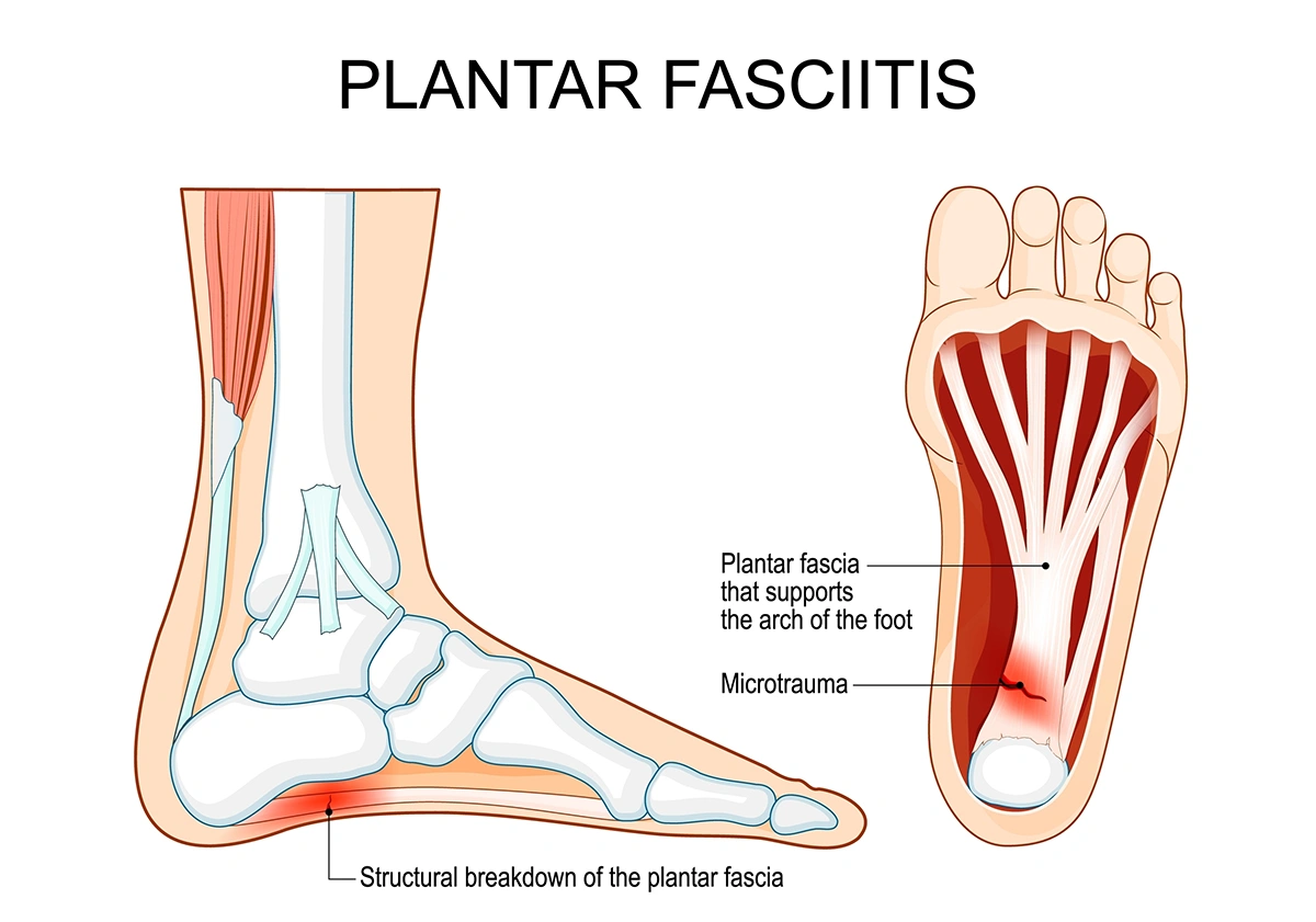
Dr Seng Chusheng is a consultant orthopaedic surgeon at Axis Orthopaedic Centre, specialising in foot and ankle surgery and restorative joint medicine. His clinical expertise includes the management of chronic plantar fascia degeneration, minimally invasive foot surgery, and advanced soft tissue repair. Over the course of his career, he has performed more than a thousand surgeries. He manages a broad range of conditions, including mechanical heel pain and sport-related lower limb injuries.
Dr Seng received advanced fellowship training at the Assal Centre in Geneva, Switzerland, focusing on complex deformities, trauma and minimally invasive techniques. Before entering private practice, he served as a consultant orthopaedic surgeon at Singapore General Hospital and continues as a visiting consultant. His dedication to patient care has been recognised with the SingHealth Service With A Heart Award. Actively involved in research and education, Dr Seng regularly presents at local and international conferences and has published extensively in peer-reviewed journals, reflecting his commitment to advancing orthopaedic practice.
Dr Seng received advanced fellowship training at the Assal Centre in Geneva, Switzerland, focusing on complex deformities, trauma and minimally invasive techniques. Before entering private practice, he served as a consultant orthopaedic surgeon at Singapore General Hospital and continues as a visiting consultant. His dedication to patient care has been recognised with the SingHealth Service With A Heart Award. Actively involved in research and education, Dr Seng regularly presents at local and international conferences and has published extensively in peer-reviewed journals, reflecting his commitment to advancing orthopaedic practice.


