
Medial Collateral Ligament (MCL) Tear: How Is It Treated?
At Axis Orthopaedic Centre, our fellowship-trained orthopaedic surgeon Dr Seng Chusheng offers personalised treatment plans that are tailored to the severity of each MCL injury, whether through non-surgical management or surgical repair.
What is the MCL?
The medial collateral ligament is one of the key ligaments stabilising the knee joint. It runs along the inner side of the knee, connecting the femur (thighbone) to the tibia (shinbone). The MCL helps resist forces that push the knee inward, which is why it is particularly vulnerable during twisting motions or direct blows to the outside of the knee.
How Does an MCL Tear Happen?
An MCL tear occurs when the ligament on the inner side of the knee is overstretched or torn due to excessive force. This is often due to:
- A sudden twisting of the knee during sports
- A direct impact on the outer side of the knee
- Falls or trauma that stress the inside of the knee joint
Symptoms of an MCL Tear
MCL tear symptoms vary depending on the tear's severity. Common signs include:
- Pain on the inner side of the knee
- Swelling and tenderness
- Instability or the knee giving way
- Difficulty straightening or bending the knee
How Are MCL Tears Diagnosed?
Orthopaedic surgeons often begin with a thorough physical examination, assessing the stability of the knee and pinpointing the area of pain.
Diagnostic tools include:
- Magnetic Resonance Imaging (MRI): MRI scans provide detailed visualisation of soft tissues, helping to determine the extent of MCL damage and identify any concurrent injuries, such as ACL tears or meniscal pathology.
- X-rays: While MCL injuries do not involve bone, X-rays may be performed to rule out associated fractures or avulsion injuries where a ligament may pull a small fragment of bone away.
Grading of MCL Tears
MCL injuries are classified into three grades based on the extent of the ligament damage and the resulting joint instability. Accurate grading helps guide appropriate treatment and predict recovery time.
- Grade I (Mild): Involves microscopic tearing of the ligament fibres without loss of structural integrity. The knee remains stable, and symptoms are typically limited to mild pain and tenderness along the inner knee.
- Grade II (Moderate): Represents a partial tear of the ligament. Patients may experience noticeable discomfort, localised swelling, and mild to moderate instability during movement or weight-bearing activities.
- Grade III (Severe): Involves a complete rupture of the MCL. This results in significant pain, swelling, and marked instability of the knee joint, often making it difficult to bear weight or maintain balance. Grade III tears often involve injuries to other ligaments like the ACL, which influences treatment planning.
Treatment Options
Treatment options range from non-surgical methods to surgical intervention, depending on the tear's severity.
Conservative Management (Grade I and II):
Most Grade I and II MCL tears respond well to non-surgical treatment, owing to the ligament's rich blood supply, which supports natural healing. Key components of conservative treatment include:
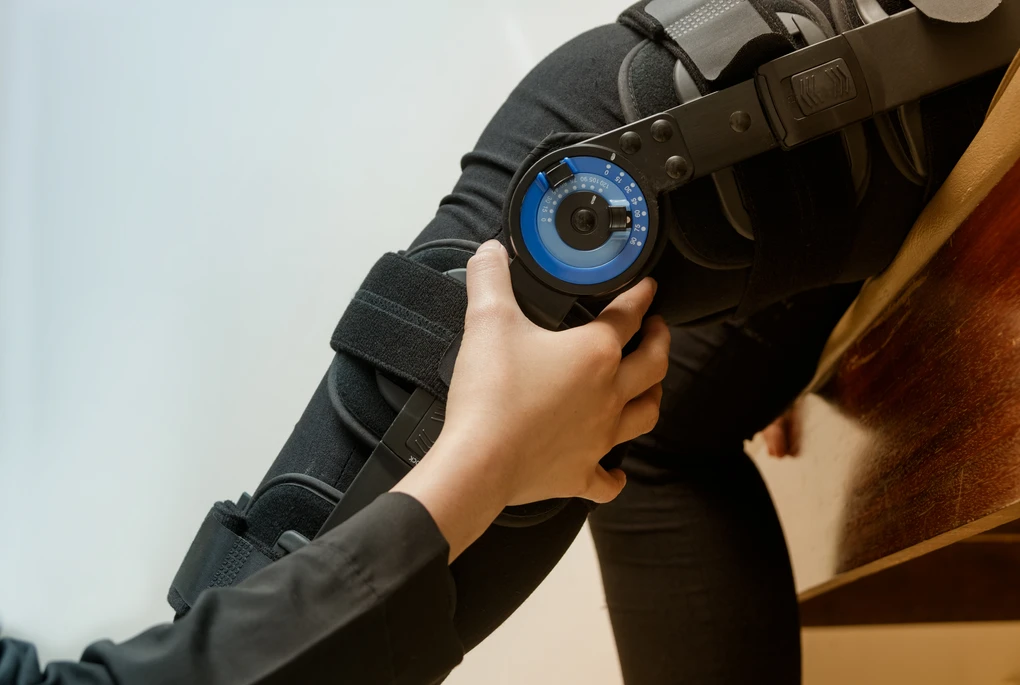
- Rest and Bracing: Activity modification and the use of a hinged knee brace help stabilise the joint and prevent further stress during the healing phase.
- Physiotherapy: A personalised rehabilitation programme that focuses on restoring range of motion, muscle strength, and proprioception to support a safe return to activity.
- Cold Therapy and Medications: Ice application and non-steroidal anti-inflammatory drugs (NSAIDs) are used to reduce swelling and alleviate discomfort.
With proper adherence to the rehabilitation programme, most patients return to sport within 4 to 12 weeks, depending on the severity.
Surgical Treatment (Grade III or Multi-ligament Injuries)
Surgical intervention for MCL tears is typically reserved for Grade III injuries, multi-ligament damage, or cases where the MCL does not heal adequately with non-surgical treatment. It is also often necessary when combined with other knee injuries, such as ACL or meniscus tears, requiring comprehensive repair.
Minimally invasive MCL repair or reconstruction can restore ligament function and knee stability. When reconstruction is needed, surgeons may use either autografts (the patient's own tissue) or allografts (donor tissue) to provide durable support. After surgery, a structured physiotherapy programme is essential to support a safe and confident return to daily activities or sports.
Recovery Timeline
The duration of recovery from an MCL injury depends on the severity of the tear and the chosen treatment approach:
- Grade I (Mild): Recovery typically takes 1 to 3 weeks with rest and rehabilitation.
- Grade II (Moderate): Healing usually occurs within 4 to 6 weeks, supported by bracing and physiotherapy.
- Grade III (Severe, Non-Surgical): Recovery may take 6 weeks or more, with a structured rehabilitation programme.
- Post-Surgical Recovery: When surgery is required, return to full activity may take 3 to 6 months, depending on the extent of associated injuries and the patient’s response to rehabilitation.
Why Consult Dr Seng Chusheng for MCL Tears?
If you are experiencing knee instability, persistent pain or swelling, or difficulty fully bending or straightening your knee, it is time to consult a specialist. Prompt diagnosis and treatment are key to preventing chronic issues and protecting your long-term joint health.
At Axis Orthopaedic Centre, Dr Seng offers expert care rooted in his specialised experience in sports injuries and ligament reconstruction. Patients benefit from:
- Advanced diagnostics
- Minimally invasive treatment options
- Personalised rehabilitation plans
With a patient-first approach, Dr Seng is committed to helping you regain strength, stability, and confidence in your knee so you can return to the activities you love.

Singapore 329563
View Us On Google Map

For foreign patients, tele-consult on zoom or WhatsApp can be done if pre-arranged in advance. Please email or WhatsApp first to enquire.
